
Digital X-Rays in Lakewood, CO
Digital X-rays in Lakewood, Colorado, allow Dr. Dian Li to see detailed images of your teeth and bone with minimal radiation exposure. This fast, safe technology helps detect problems early and plan treatment precisely.
Digital X-Rays Explained
Digital dental X-rays use an electronic sensor to capture images of your teeth and jaws. The sensor sends the image to a computer almost instantly, allowing the dental team to view and adjust contrast or magnification as needed. Compared with traditional film, digital imaging uses a lower radiation dose and eliminates chemical processing. The result is fast, clear pictures that support accurate diagnosis and treatment planning.
Digital radiographs are an important part of preventive and restorative care. They reveal decay that may not be visible during a visual exam, show the health of tooth roots, and help evaluate bone levels around teeth. They are also used to plan treatments such as fillings, root canals, implants, and orthodontics.
Common Image Types
- Bitewing images show the upper and lower back teeth to check for cavities and bone levels.
- Periapical images focus on one or two teeth from crown to root to assess the root, nerve, and supporting bone.
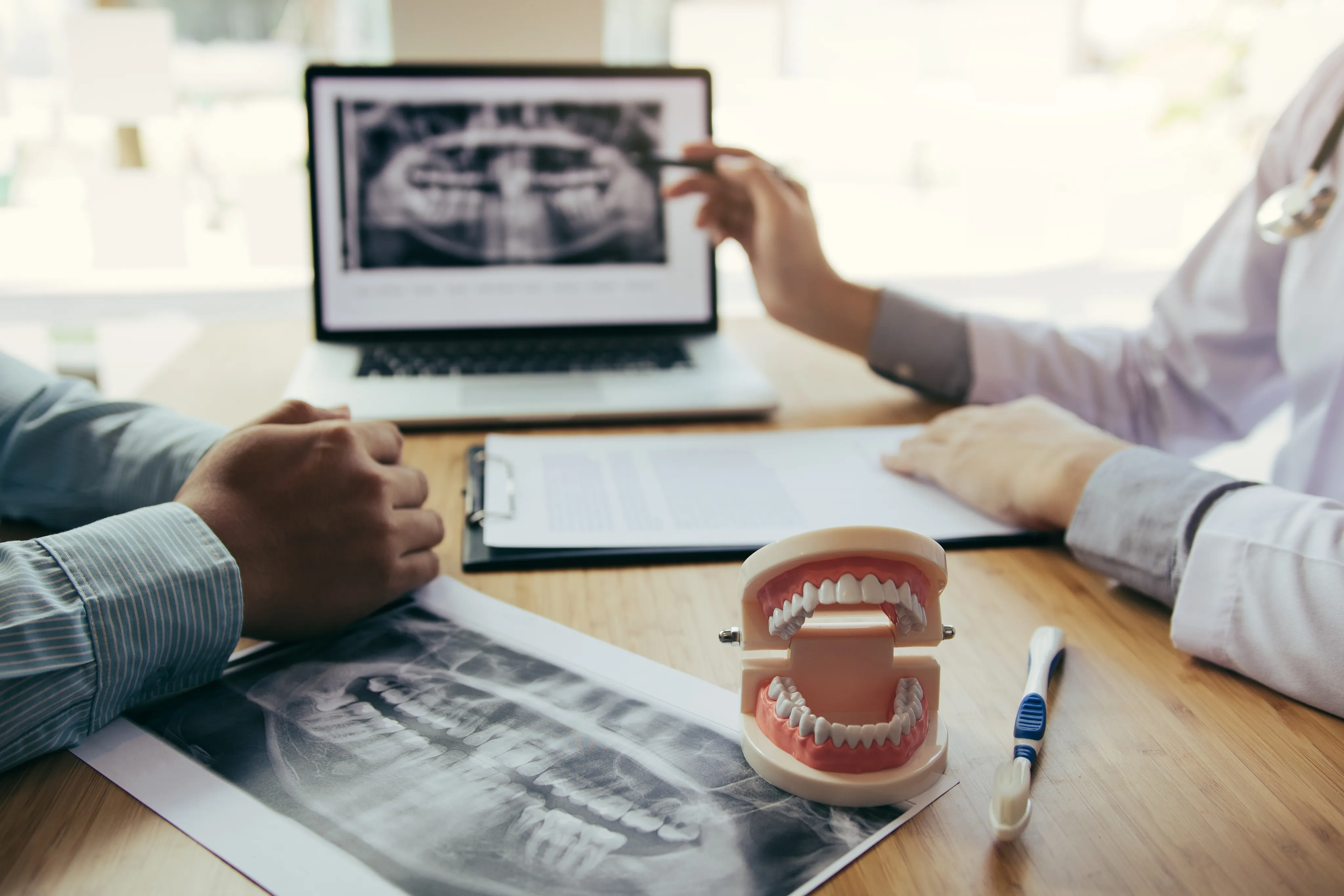
- Panoramic images capture the entire mouth, including jaws and sinuses, to evaluate growth, impacted teeth, and joint structures.
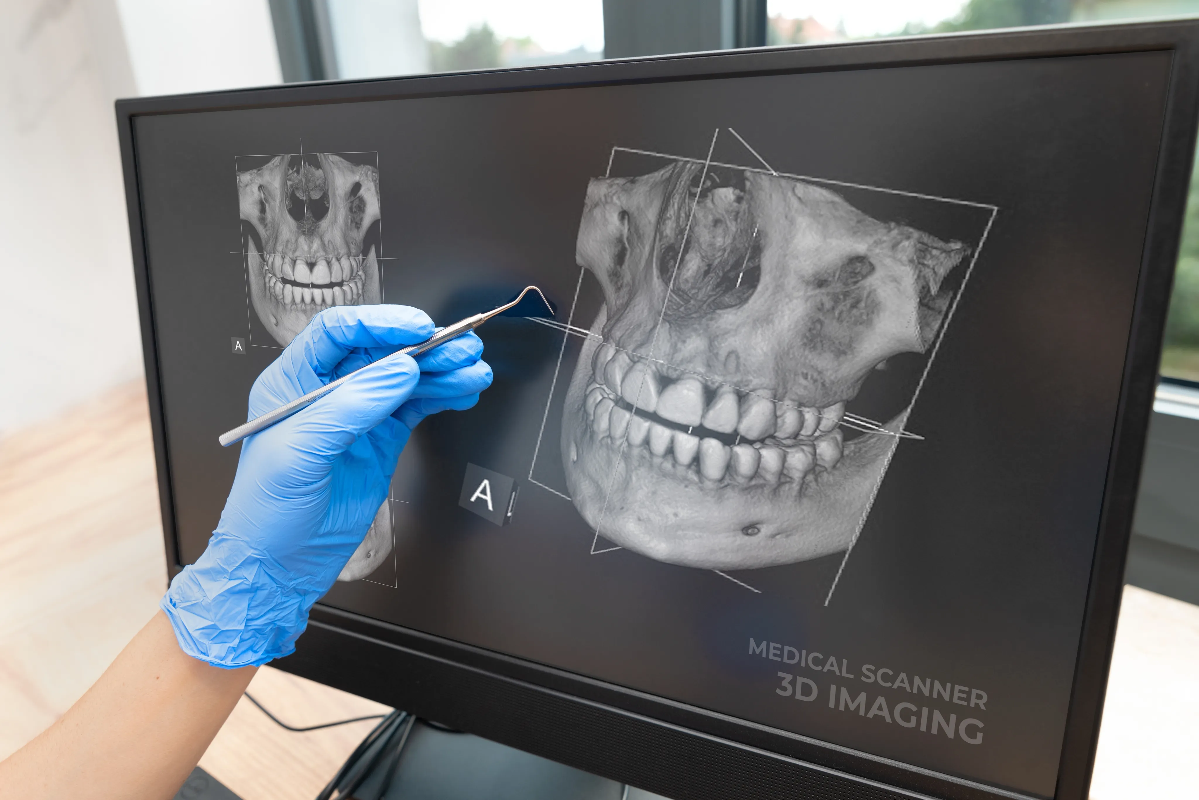
- Cone beam CT may be used for 3D views when detailed planning is needed, such as implants or complex cases.
Benefits of Digital X-Rays
Digital technology improves safety, speed, and clarity. It also supports better communication, since images can be displayed chairside and explained in real time. Key advantages include:
- Lower radiation dose compared with traditional film systems.
- Immediate image preview that helps reduce time in the chair.
- High-resolution detail that supports precise diagnosis.
- Eco-friendly workflow without chemical developers.
- Secure digital storage for easy comparison over time.
How Digital X-Rays Work
The process is quick and comfortable. A small sensor or plate is positioned inside your mouth or a camera rotates around your head for a panoramic image. A brief exposure captures the picture, which appears on a monitor within seconds. Our dentist reviews the images with you, pointing out findings and discussing next steps if treatment is needed.
- Sensor placement ensures the right area is captured with minimal retakes.
- Protective measures such as a lead apron and thyroid collar are used when appropriate.
- Real-time review allows adjustments or additional angles if more detail is required.
- Images are saved securely for future comparison and monitoring.
What to Expect
Most patients find digital X-rays comfortable, with only brief pressure from the sensor. The number and type of images depend on your needs. New patients often receive a baseline set, while routine checkups may include bitewings to monitor for decay. If you are pregnant or think you might be, let the team know so protective steps can be taken or timing adjusted. Radiation exposure from digital dental X-rays is low and is kept as minimal as reasonably achievable while still gathering the information needed for safe care.
Frequently Asked Questions
Yes. Digital X-rays use significantly less radiation than traditional film. Protective measures such as lead aprons and thyroid collars add further safety. Images are taken only when clinically needed.
Frequency depends on your oral health, risk for decay, and treatment history. Many patients receive bitewing images once a year and a full set every three to five years. Your dentist adjusts the schedule based on your needs.
X-rays reveal many issues such as hidden decay, bone loss, and infections, but they do not show everything. A clinical exam, intraoral camera images, and other diagnostic tools work together for a complete evaluation.
Yes, when clinically indicated. X-rays help monitor developing teeth, check for decay between baby teeth, and assess jaw growth. The radiation dose is very low with digital sensors.
Your dentist will explain the finding, discuss treatment options, and help you understand next steps. Early detection often means simpler, less invasive treatment.
You have the right to decline, but X-rays provide essential information that may not be available through a visual exam alone. Your dentist will explain why images are recommended and how they support your care.
Safe, Fast Dental Imaging
Contact Belmar Smiles at 303-935-3574 to schedule your appointment with digital X-ray imaging included.